Products
ImageCyte products combine coverslip glass with precision-engineered micro-features to deliver clearer images, more consistent experiments, and higher throughput—without disrupting existing workflows. Designed to work seamlessly with standard microscopes and automation, our plates reduce manual handling, minimize variability, and unlock reliable single-cell, 3D, and model-organism imaging at scale. The result is better data, faster decisions, and experiments you can trust.
SAMPLES AVAILABLE NOW
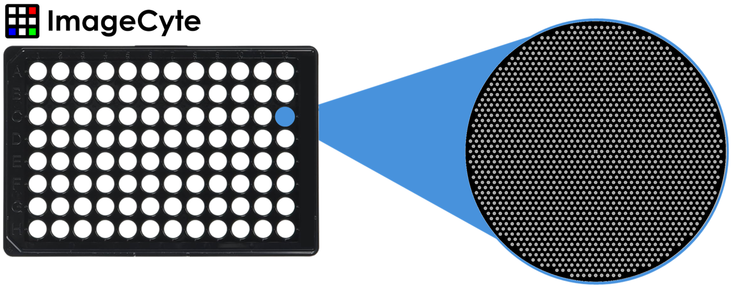
Single-cell imaging
High-density nanowell plates designed to physically isolate individual cells for high-content imaging in suspension or weakly adherent conditions. Glass-bottom wells enable high-NA imaging while maintaining cell position during washing, staining, and longitudinal experiments. These plates support scalable single-cell phenotypic screening with improved reproducibility.

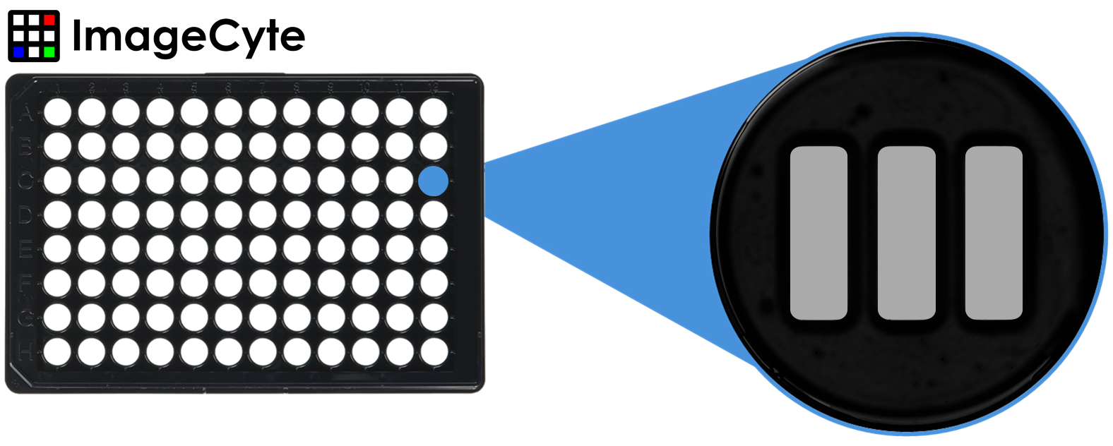
Multi-cell and clonal expansion
Nanowell formats optimized for studying cell–cell interactions, small multicellular assemblies, and clonal outgrowth. Defined well geometry constrains cell populations without chemical modification, enabling consistent imaging of colony formation and interaction dynamics. Ideal for workflows requiring controlled multicellular organization at scale.
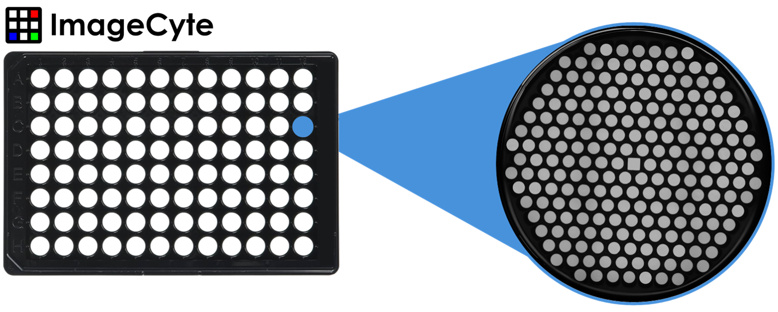
Spheroid Screening
Glass-bottom nanowell plates designed for the formation, maintenance, and high-content imaging of uniform spheroids. Defined well geometry promotes consistent spheroid size while preventing aggregation and drift during media exchange. These plates enable scalable, reproducible spheroid assays with high optical quality.
Organoid Screening
Glass-bottom nanowell plates engineered to support parallel imaging of thousands of organoids in a single experiment. Physical confinement prevents organoid collision and aggregation, improving imaging consistency and assay reproducibility. These plates enable high-throughput organoid screening without compromising optical quality.

Zebrafish Screening
Plates that passively align zebrafish embryos for rapid, reproducible imaging, eliminating the need for agarose-based mounting in many workflows. Precision nanowell geometry standardizes orientation across wells, reducing manual handling and variability. Designed for high-throughput developmental and screening studies using standard microscopy platforms.